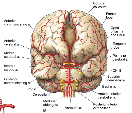
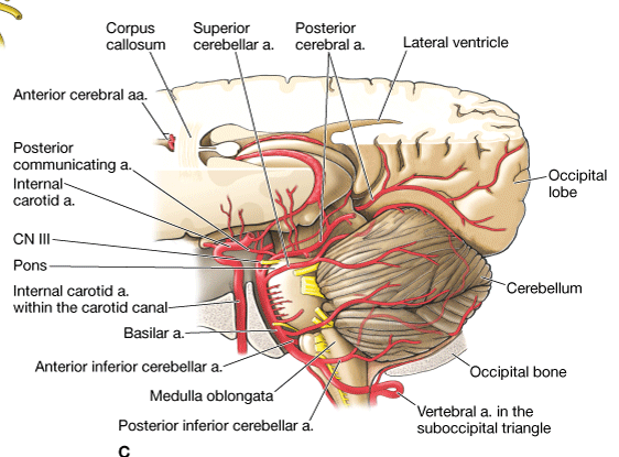
Vertebral Arteries
Each vertebral artery arises from the subclavian artery and ascends through the transverse foramina of C1–C6. The vertebral artery courses horizontally across C1 through the suboccipital triangle before entering the skull via the foramen magnum (Figure 16-3A–C). After penetrating the dura mater, the vertebral arteries then course along the inferior aspect of the medulla oblongata before converging into the basilar artery on the pons. Branches of the vertebral arteries travel to the spinal cord, the meninges, and the brainstem. The major branches are as follows:
- Posterior inferior cerebellar arteries. Course between the origins of cranial nerve (CN) X and CN XI en route to the inferior surface of the cerebellum. Often referred to by the acronym PICA.
- Posterior cerebral arteries. The terminal branches of the basilar artery provide vascular supply to that part of the brain base that is superior to the tentorium cerebelli. CN III and CN IV exit the brain between the superior cerebellar and the posterior cerebral arteries.
Basilar Artery
The basilar artery ascends along the ventral surface of the pons and gives rise to the following branches:
- Anterior inferior cerebellar artery. Courses along the inferior surface of the cerebellum.
- Superior cerebellar artery. Courses along the superior surface of the cerebellum.
Circle of Willis
The anterior communicating artery connects the two anterior cerebral arteries, and the posterior communicating arteries connect the internal carotid and posterior cerebral arteries. As a result of these connections, an arterial circle, known as the cerebral arterial circle (of Willis), is formed around the infundibulum (stalk connecting the pituitary gland to the hypothalamus).
 The structure of the arterial supply of the brain into the circle of Willis provides a collateral circulatory pathway in the cerebral circulation. As a result, if one part of the circle becomes narrowed or blocked or if one of the four arteries supplying the circle is narrowed or blocked, blood flow from the other vessels preserves cerebral perfusion. This collateral circulation is made possible by the lack of valves in arteries. However, some of the numerous arteries arising from this circle that penetrate the brain substance are small and are considered end arteries (without collateral circulation). Therefore, if an end artery becomes narrowed or blocked, ischemia may occur in the region of the brain that is uniquely supplied by that end artery.